Treating Our Patients Like Family
A Bright, Beautiful Smile Says Many Things: Confidence, Happiness, and Overall Health.
Office Hours
Your Dentist in San Antonio, TX
Emergency Care
TREAT YOU LIKE FAMILY
ALL UNDER ONE ROOF
HERE FOR YOU!
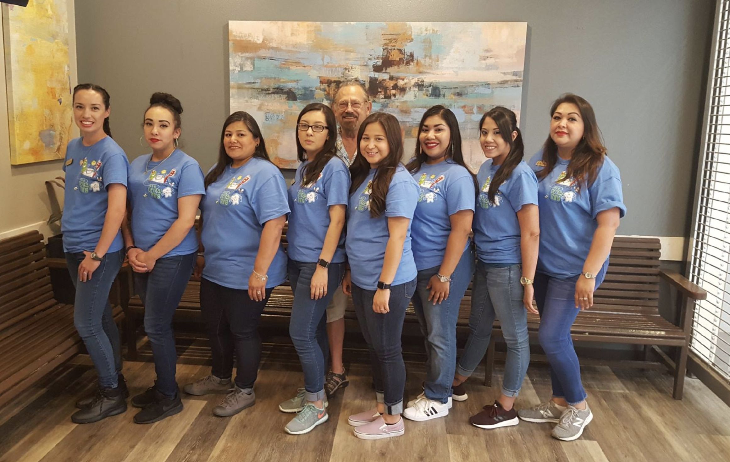
Our Patients: Like Family, Every Time, All Of The Time
Our offices are a home away from home for each of our staff members. Sometimes we see and work with each other more than we see our own families, wo we strive to make each office like our own home. Each patient is welcomed into our home as a long-lost family member. Our main goal is to provide quality care with the bedside manner that is deserving of our loved ones. We invite you to join our practice and be a part of our dental family.
We have several state-of-the-art locations for your convenience, with dental experts on staff to provide specialized care.


Dental Emergency?
New Patient? First Visit?
Our goal is to provide you with the highest level of dental care available today. Thank you for your confidence in our dental office and we look forward to meeting you.
How Can We Help You?
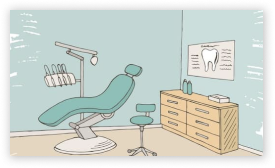
Time for a checkup?
We’ve got you covered with personalized cleanings, painless fillings, crowns, dentures, and bridges.
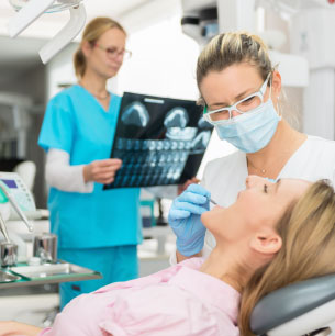
Fix damaged teeth?
Expert surgical care from the team you know and trust. Implants, root canals, extractions and more.
Improve your smile?
We’ll help you understand high-tech cosmetic options, like Invisalign, veneers, and teeth whitening.
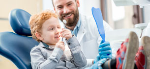
Get to know us?
Led by Dr. Shiva Izzadoust, our team is proud to offer a fresh take on going to the dentist in San Antonio.
Most Commonly Asked
Questions on Google:
Our Locations
Westover Hills Family
Dental
Ste. 290 San Antonio, TX 78251